Disulfide Bond Formation in Bacterial Cell-Free Systems
Cell-free systems need multiple layers to form disulfide bonds correctly.

Bacterial cell-free protein synthesis has a redox problem, and it has one because E. coli itself has a redox preference. The cytoplasm of a living E. coli cell runs strongly reducing, and that same chemistry carries straight into a cell extract unless someone intervenes. Getting disulfide bonds to form correctly in that extract takes a specific, layered set of interventions, and understanding how each one works, and where each one falls short on its own, is the difference between a folded protein and a tube of aggregate.
The reducing cytoplasm isn't a design flaw. It's protective. Most of E. coli's metabolic enzymes depend on free thiols to function, and an oxidizing cytoplasm would wreck them. Two enzyme systems do the protecting: thioredoxin reductase (TrxB) and glutathione reductase (Gor), key components of the thioredoxin and glutaredoxin/glutathione pathways respectively. Both actively break disulfide bonds or stop them from forming in the first place. Drop a protein that needs paired cysteines into an unmodified extract, and those cysteines are likely to stay as free thiols, or worse, pair up with the wrong partner and lock the chain into a misfolded, insoluble mess.
E. coli does make disulfide bonds, just not in the cytoplasm. It happens in the periplasm, where DsbA, a thioredoxin-family oxidase with a Cys-X-X-Cys active site, introduces bonds directly, and DsbB regenerates DsbA by handing electrons off to membrane quinones. When DsbA gets it wrong and pairs the wrong cysteines, DsbC and DsbD step in to isomerize the mismatch into the correct pairing, with glutathione lending a hand in keeping the periplasm's redox balance steady. This periplasmic system is effectively the blueprint that cell-free engineers are trying to rebuild inside a test tube.
None of this is a side curiosity. Antibodies, cytokines, hormones, proteases, and lectins, some of the most valuable protein classes in medicine, nearly all depend on correctly formed disulfide bonds to fold and function. Solving this problem in a cell-free format has direct, immediate stakes for how these molecules get made.
What cell-free protein synthesis is and why its open architecture matters for redox chemistry
Cell-free protein synthesis, or CFPS, pulls transcription and translation out of a living cell and runs them in an open reaction. A cell extract, mostly from E. coli in the bacterial case, gets combined with a DNA or mRNA template, amino acids, an energy source, and cofactors, and the mix produces protein without any cell membrane standing in the way.
That openness is the whole argument. There's no viability to protect and no membrane to work around, so the researcher can reach directly into the reaction chemistry. Add an oxidant. Titrate the glutathione ratio. Drop in a purified isomerase. Shift the pH. None of that is possible inside an intact bacterium, where changing the internal environment risks killing the cell before it finishes making the protein. Goerke and Swartz laid this out plainly in Biotechnology and Bioengineering in 2008: the open reaction environment of a cell-free system lets researchers push the conditions needed for disulfide-bonded proteins to fold, and do it fast and at reasonable cost.
Compare that to the cell-based alternatives. Periplasmic secretion in E. coli can work, but it's capped by secretion efficiency and by how large the target protein is. Mammalian and insect expression systems, CHO cells for instance, handle disulfide bonds as a matter of course, but they're slow, expensive to run, and awkward to adapt for screening at any real scale. CFPS isn't a workaround for when those systems fail. For disulfide-bonded targets, the control it offers over the reaction chemistry makes it a legitimately better starting point.
That's the design space OpenCFPS™ systems built on E. coli extract sit in: plate-compatible, built for automation, and quality-controlled lot to lot, so the redox starting point is a known quantity before any intervention gets layered on.
The redox buffer: setting glutathione ratios to create an oxidizing environment
The first lever is chemical, not genetic. A glutathione redox buffer, a defined mix of oxidized glutathione (GSSG) and reduced glutathione (GSH), gets added straight into the reaction to push the thiol equilibrium toward oxidation. What sets the redox potential isn't the total glutathione concentration. It's the ratio of GSSG to GSH, and tuning that ratio is how the reaction mimics the oxidizing chemistry of the natural periplasm.
Seki et al., in a 2019 bioRxiv preprint, laid out a working formulation: 2 mM GSSG added alongside 1.2 mM ATP, 130 mM potassium glutamate, and 12 mM magnesium glutamate, run in 384-well black-walled flat-bottom plates.
But the buffer alone doesn't hold. In an untouched extract, endogenous glutathione reductase keeps converting that GSSG back into GSH, and the oxidizing potential collapses almost as fast as it's set up. Adding the buffer without dealing with Gor is like propping a door open against a strong spring: it swings shut the moment the pressure's removed. That's exactly why extract engineering and iodoacetamide treatment aren't optional extras. They're what makes the redox buffer actually stick.
The broader literature backs this up directly: glutathione buffer paired with a disulfide isomerase such as DsbC has been shown to support active, correctly folded protein production in cell-free reactions.
Disabling endogenous reductases: strain engineering and iodoacetamide pretreatment
Two enzymes need to be shut down for an oxidizing buffer to hold: Gor and TrxB.
One route is genetic. Deleting or inactivating gor in the extract's source strain cuts reductase activity substantially, though not to zero. The strain KGK10 carries that deletion, and it also carries a tagged trxB allele, which lets TrxB be pulled out of the extract by affinity purification after lysis. That matters because in an unmodified strain, TrxB doesn't just reduce disulfide bonds directly, it also works against the oxidizing buffer through its reductase activity.
The other route is chemical: iodoacetamide, or IAM, added before the reaction starts. IAM alkylates free sulfhydryl groups on the endogenous reductases, and that alkylation is irreversible, so the enzymes stay off for the life of the reaction. US Patent 7,871,794 (Knapp et al., issued 2011) puts the working range at 5 to 500 μM, with 20 to 75 μM as the effective window and 50 μM as the documented working dose.
There's a catch, though, and it's the reason the genetic and chemical routes end up paired rather than used alone. At higher IAM doses, the treatment also knocks out glyceraldehyde 3-phosphate dehydrogenase, the enzyme that lets the reaction run on glucose for energy. Knock that out, and the energy source goes with it. Starting from a Δgor background like KGK10 lowers the IAM dose needed to control the reductases, which keeps G-3PDH intact and glucose usable as fuel (Knapp et al., Biotechnology and Bioengineering, 2007).
IAM's effectiveness also isn't flat across conditions. The patent data show its ability to stabilize the redox environment holds most reliably in a moderate pH range, and drifting to either extreme changes how much is needed to get the same effect.
The practical rule that falls out of all this: genetic knockout lowers the IAM burden and protects the energy pathway, but IAM still has a job to do even in an engineered strain, mopping up whatever reductase activity the deletion didn't catch.
Why isomerization, not oxidation, is typically the yield-limiting step
Getting the environment oxidizing enough is only half the job, and arguably not the harder half.
DsbA doesn't pick its partners. It oxidizes cysteines roughly in the order it encounters them, not by matching correct pairs, which means a protein with more than one disulfide bond will, purely by chance, form some wrong pairings even when the surrounding chemistry is fully oxidizing. The more cysteines involved, the worse the odds get.
Ryabova et al., writing in Nature Biotechnology in 1997, ran the experiment that pins this down. Adding protein disulfide isomerase (PDI) to the reaction tripled yield over a glutathione redox system running alone. Adding more DsbA did nothing of the sort. That result reframes the whole problem: the bottleneck isn't whether bonds form, it's whether the wrong ones get corrected.
Once that's established, the fix for low yield stops being "add more oxidant." Past a certain point, more GSSG or more DsbA doesn't move the needle, because the constraint has already shifted downstream to the proofreading step. Ryabova's group also tested DnaK and DnaJ chaperones and found they raised the amount of soluble protein without raising the amount of functional protein, which is its own kind of evidence: the ceiling isn't aggregation. It's whether the disulfides land in the right place.
Adding disulfide isomerases: DsbC, PDI, and FkpA as active folding catalysts
DsbC is the periplasmic isomerase built for exactly this job. It catalyzes a thermodynamically driven reshuffle of incorrectly paired disulfides, nudging the protein toward whatever pairing sits at the lowest energy state, which in a properly folded protein is usually the native one. Seki et al. confirmed DsbC does this inside the cell-free reaction itself, not just in isolation, and it turned out to matter for one of the harder benchmark targets in the field: urokinase, a protease with nine disulfide bonds, reached 40 μg/mL of active protein using a glutathione redox buffer, DsbC, and pH optimization together, in a plain batch reaction.
PDI, the eukaryotic version of the same idea, works as both an isomerase and a chaperone, and it's what drove the threefold yield gain Ryabova's group reported. That a eukaryotic enzyme functions inside a bacterial cell-free reaction says something useful on its own: isomerase activity isn't locked to its organism of origin, it can be imported.
FkpA covers a different fold entirely. It's a peptidyl-prolyl cis-trans isomerase, and it matters most for immunoglobulin-like structures, where proline isomerization and disulfide formation are separate jobs that both need doing for the chain to assemble correctly. Sutro Biopharma's work identified DsbC and FkpA together as the two factors needed to get immunoglobulins expressed at high yield in a cell-free system. Sutro's own patent, US 10,190,145 (Yam et al., 2019), describes the combination of a disulfide isomerase and a prolyl isomerase as producing a synergistic gain in correctly folded, active protein, not just an additive one. The patent's dose-response numbers make the point concrete: raising DsbC from 0 to 6.4 μM with no PDI present takes activity from 0 to roughly 160 units, and adding just 0.5 μM PDI on top of that 6.4 μM DsbC pushes it to around 180. DsbC does the heavy lifting at low doses; PDI adds a smaller but real gain once DsbC is near saturation.
A broader chaperone set, DnaK, DnaJ, GroEL, GroES, gets used alongside these isomerases to keep proteins from aggregating during folding. They're a complement to the isomerase work, not a substitute for it, which lines up with what Ryabova's chaperone data already showed. And isomerases don't have to be added as purified protein each time. They can also be built into the source strain and expressed directly into the extract during preparation, which is the approach taken by certain engineered source strains.
SHuffle and engineered source strains as a simpler path to isomerase-competent extracts
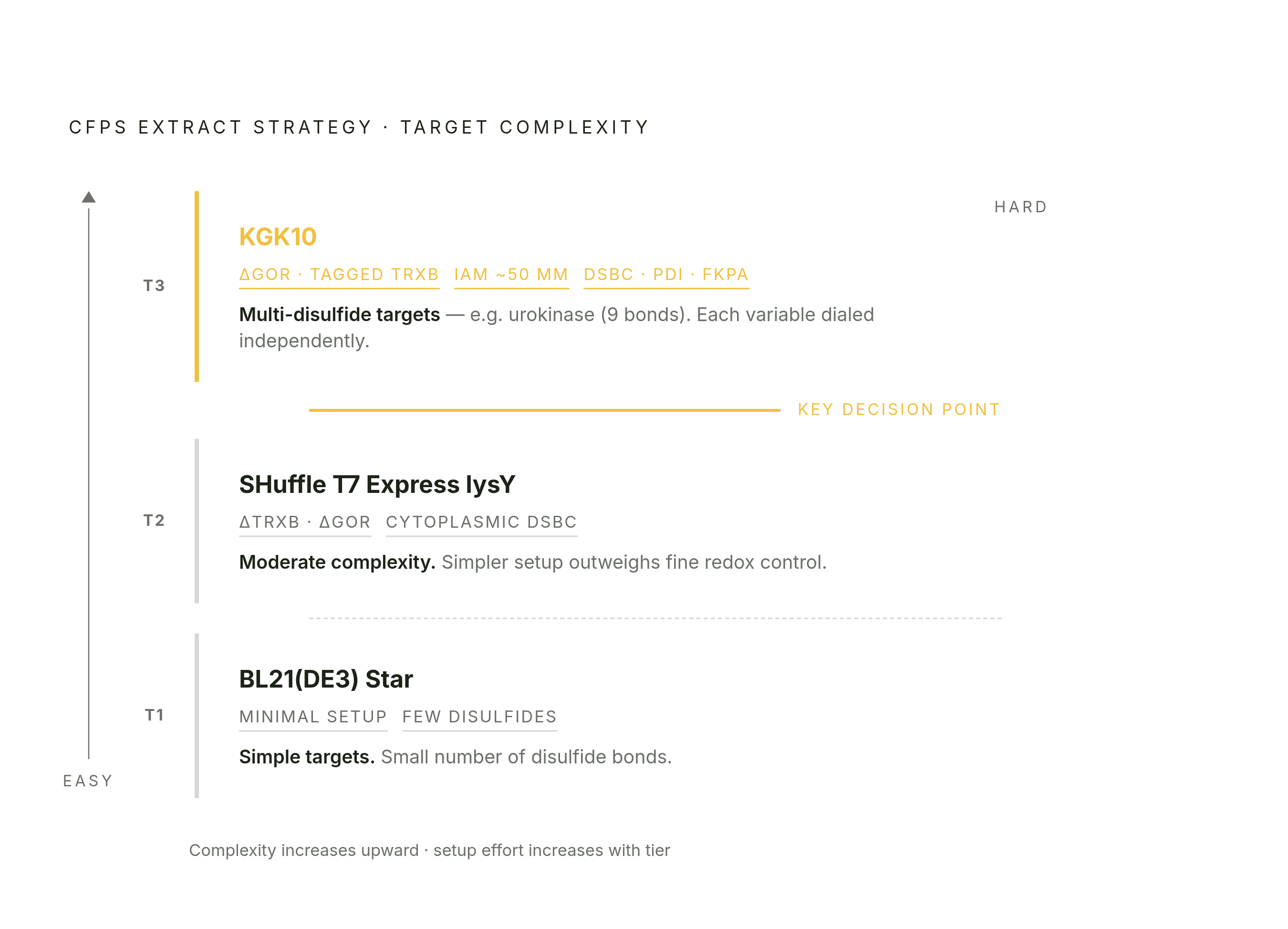
SHuffle T7 Express lysY is a commercial E. coli strain that folds both fixes into one background: it expresses DsbC directly in the cytoplasm, and it carries deletions in both trxB and gor. One strain handles the reductase problem and supplies isomerization capacity at the same time.
Using a SHuffle-derived extract means DsbC doesn't need to be added separately, and the IAM dose needed on top of it drops, though it isn't necessarily eliminated. The appeal of this approach is that the strain has already done the engineering work before the extract is even made, potentially reducing the need for supplemental enzymes. Hunt et al., in a 2025 methodological review in Chemical Reviews, point to strains engineered for cytoplasmic disulfide formation as useful for producing full-length antibodies in cell-free reactions.
None of the three approaches, IAM plus KGK10, SHuffle on its own, or plain BL21(DE3) Star, is a universal answer. SHuffle extract has its own productivity profile, and a target that expresses well in a KGK10-plus-exogenous-DsbC setup won't necessarily behave the same way in SHuffle, or vice versa. BL21(DE3) Star, for its part, has been used directly to express disulfide-bonded proteins, but the literature is consistent that it holds up only for targets with a small number of bonds, not the more demanding multi-disulfide cases.
The practical decision comes down to target complexity against setup effort. BL21 fits simple targets, or cases where extract availability is the real constraint. SHuffle fits moderately complex targets when a simpler reaction setup is worth more than fine control. KGK10-derived extract with IAM and exogenous isomerases fits the hardest targets, where each variable, redox potential, reductase activity, isomerase dose, needs to be dialed in on its own.
What these strategies have achieved: model proteins that benchmark the system
Urokinase is the benchmark that carries the most weight, given nine disulfide bonds is a demanding target by any measure. Glutathione redox buffer, DsbC, and pH optimization together produced 40 μg/mL of active protease in a simple batch reaction, with activity confirmed rather than just a folding proxy.
BPTI and lysozyme C came out of the same body of work at milligram quantities, both catalytically active and correctly folded, and both made as ¹⁵N-labeled protein, which matters beyond the folding question itself: isotope labeling for NMR structural work is one of CFPS's clearest advantages over cell-based expression, since feeding a labeled amino acid into an open reaction is far simpler than getting a living cell to take it up cleanly.
scFv antibody fragments reached 230 μg/mL under generic CFPS conditions in Goerke and Swartz's 2008 work, and the same platform produced a therapeutic protein at 710 μg/mL and a vaccine fusion protein at 300 μg/mL, evidence the approach isn't narrowly tuned to antibody fragments alone.
The open reaction format also simplifies downstream characterization, since expressed protein can in principle move directly into binding or activity assays., which shows something practical: a correctly folded, disulfide-bonded protein made this way can move directly into downstream assay work, cutting out a purification step that would otherwise sit between expression and any usable result.
And the industrial end of this isn't theoretical. Sutro Biopharma's OCFS platform runs coupled transcription-translation at reaction volumes spanning from bench scale to tens of thousands of liters, with disulfide and prolyl isomerases supplemented into the mix, and expressing heavy and light antibody chains separately to improve how well the final IgG assembles. That's the clearest evidence available that the bacterial CFPS approach, redox buffer, reductase control, and isomerase supplementation together, holds up well past the scale of a lab bench.
Sources
- 7871794
- Development of cell-free protein synthesis platforms for disulfide bonded proteins | Request PDF
- Cell-free synthesis of proteins that require disulfide bonds using glucose as an energy source | Request PDF
- 10190145
- Cell-Free Gene Expression: Methods and Applications
- analyticalsciencejournals.onlinelibrary.wiley.com
- nature.com
- biorxiv.org